Home
/ Structure Of Plant Cell Under Electron Microscope / Cell And Organelles Dr Jastrow S Electron Microscopic Atlas : Electron microscope, microscope that attains extremely high resolution using an electron beam instead of a beam of light to illuminate the object of study.
Structure Of Plant Cell Under Electron Microscope / Cell And Organelles Dr Jastrow S Electron Microscopic Atlas : Electron microscope, microscope that attains extremely high resolution using an electron beam instead of a beam of light to illuminate the object of study.
Structure Of Plant Cell Under Electron Microscope / Cell And Organelles Dr Jastrow S Electron Microscopic Atlas : Electron microscope, microscope that attains extremely high resolution using an electron beam instead of a beam of light to illuminate the object of study.. Essentially, an atom is the smallest unit of an element that retains the properties of the same element (iron, copper, carbon etc). Electron microscopy gives a much higher resolution showing greatly detailed cell structure. The structure of a cell as seen under an electron microscope is called ultrastructure. This means that divided further, its components (electrons, protons, and neutrons) do not retain the properties of the element. Just under the rigid cell wall is the more fluid cell membrane.
The modern understanding of cellular substructure began with the use of the electron microscope. Light passes from a bulb under the stage, through a … Essentially, an atom is the smallest unit of an element that retains the properties of the same element (iron, copper, carbon etc). Apart from the cell wall, there are other organelles that are associated with different cellular activities. The cytoplasm enclosed within the cell membrane does not exhibit much structure when viewed by electron microscopy.
What Are The Differences Between A Plant Cell And An Animal Cell from www.microscopemaster.com Certain structures can be seen only under an electron microscope. The explosion of knowledge brought about by improvements in microscopy, biochemistry, and genetics. Observation of euglena under more powerful electron microscopes have revealed the presence of an ornamented pellicle under the plasma membrane. The plant cell is surrounded by a cell wall which is involved in providing shape to the plant cell. This means that divided further, its components (electrons, protons, and neutrons) do not retain the properties of the element. Electron microscopy gives a much higher resolution showing greatly detailed cell structure. Light passes from a bulb under the stage, through a … The structure of a cell as seen under an electron microscope is called ultrastructure.
The plant cell is surrounded by a cell wall which is involved in providing shape to the plant cell.
The plant cell is surrounded by a cell wall which is involved in providing shape to the plant cell. The cytoplasm enclosed within the cell membrane does not exhibit much structure when viewed by electron microscopy. Fundamental research by many physicists in the first quarter of the 20th century suggested that cathode rays (i.e., electrons) might be used in some way to increase microscope resolution. Plant cells are eukaryotic cells present in green plants, photosynthetic eukaryotes of the kingdom plantae.their distinctive features include primary cell walls containing cellulose, hemicelluloses and pectin, the presence of plastids with the capability to perform photosynthesis and store starch, a large vacuole that regulates turgor pressure, the absence of flagella or centrioles, except in. Electron microscope, microscope that attains extremely high resolution using an electron beam instead of a beam of light to illuminate the object of study. The modern understanding of cellular substructure began with the use of the electron microscope. The explosion of knowledge brought about by improvements in microscopy, biochemistry, and genetics. Light passes from a bulb under the stage, through a … The organelles of the organism and its cytoplasm are therefore bound by a plasma membrane that allows for easier movement. Observation of euglena under more powerful electron microscopes have revealed the presence of an ornamented pellicle under the plasma membrane. Essentially, an atom is the smallest unit of an element that retains the properties of the same element (iron, copper, carbon etc). Let us have a detailed look at the plant cell, its structure and functions of different plant cell organelles. Electron microscopy gives a much higher resolution showing greatly detailed cell structure.
Essentially, an atom is the smallest unit of an element that retains the properties of the same element (iron, copper, carbon etc). Electron microscopy gives a much higher resolution showing greatly detailed cell structure. The plant cell is surrounded by a cell wall which is involved in providing shape to the plant cell. The cytoplasm enclosed within the cell membrane does not exhibit much structure when viewed by electron microscopy. The modern understanding of cellular substructure began with the use of the electron microscope.
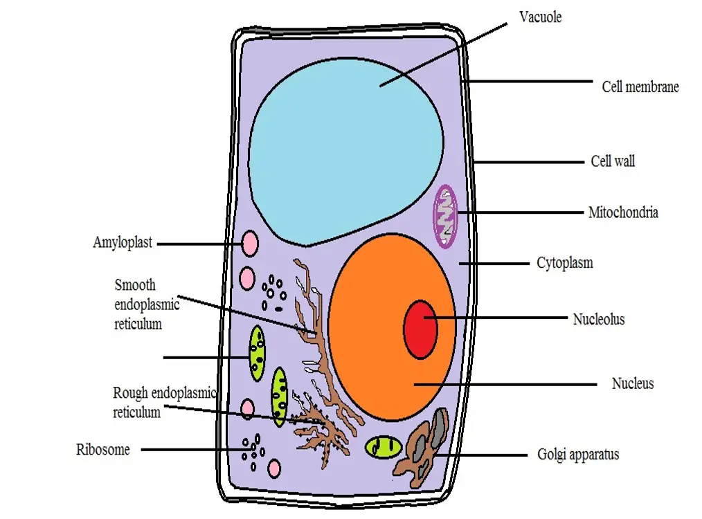
Gce Cie Biology Animal And Plant Cell Structures And Functions Biology Gce Cie Organelles Cell Function Eukaryotes from user-images.strikinglycdn.com Apart from the cell wall, there are other organelles that are associated with different cellular activities. Certain structures can be seen only under an electron microscope. Light passes from a bulb under the stage, through a … Aug 20, 2018 · structure of cell the detailed structure of a cell has been studied under compound microscope and electron microscope. The explosion of knowledge brought about by improvements in microscopy, biochemistry, and genetics. Plant cells are eukaryotic cells present in green plants, photosynthetic eukaryotes of the kingdom plantae.their distinctive features include primary cell walls containing cellulose, hemicelluloses and pectin, the presence of plastids with the capability to perform photosynthesis and store starch, a large vacuole that regulates turgor pressure, the absence of flagella or centrioles, except in. The cytoplasm enclosed within the cell membrane does not exhibit much structure when viewed by electron microscopy. Electron microscope, microscope that attains extremely high resolution using an electron beam instead of a beam of light to illuminate the object of study.
Observation of euglena under more powerful electron microscopes have revealed the presence of an ornamented pellicle under the plasma membrane.
Unlike most plant cells, this species do not have a cell wall. The structure of a cell as seen under an electron microscope is called ultrastructure. This means that divided further, its components (electrons, protons, and neutrons) do not retain the properties of the element. Electron microscope, microscope that attains extremely high resolution using an electron beam instead of a beam of light to illuminate the object of study. Let us have a detailed look at the plant cell, its structure and functions of different plant cell organelles. Certain structures can be seen only under an electron microscope. Fundamental research by many physicists in the first quarter of the 20th century suggested that cathode rays (i.e., electrons) might be used in some way to increase microscope resolution. The explosion of knowledge brought about by improvements in microscopy, biochemistry, and genetics. Atom under the microscope electron & atomic force microscopy what is an atom? The plant cell is surrounded by a cell wall which is involved in providing shape to the plant cell. Plant cells are eukaryotic cells present in green plants, photosynthetic eukaryotes of the kingdom plantae.their distinctive features include primary cell walls containing cellulose, hemicelluloses and pectin, the presence of plastids with the capability to perform photosynthesis and store starch, a large vacuole that regulates turgor pressure, the absence of flagella or centrioles, except in. Observation of euglena under more powerful electron microscopes have revealed the presence of an ornamented pellicle under the plasma membrane. Use the following animation to explore bacterial structure.
Plant cells are eukaryotic cells present in green plants, photosynthetic eukaryotes of the kingdom plantae.their distinctive features include primary cell walls containing cellulose, hemicelluloses and pectin, the presence of plastids with the capability to perform photosynthesis and store starch, a large vacuole that regulates turgor pressure, the absence of flagella or centrioles, except in. Atom under the microscope electron & atomic force microscopy what is an atom? Apart from the cell wall, there are other organelles that are associated with different cellular activities. Just under the rigid cell wall is the more fluid cell membrane. Essentially, an atom is the smallest unit of an element that retains the properties of the same element (iron, copper, carbon etc).
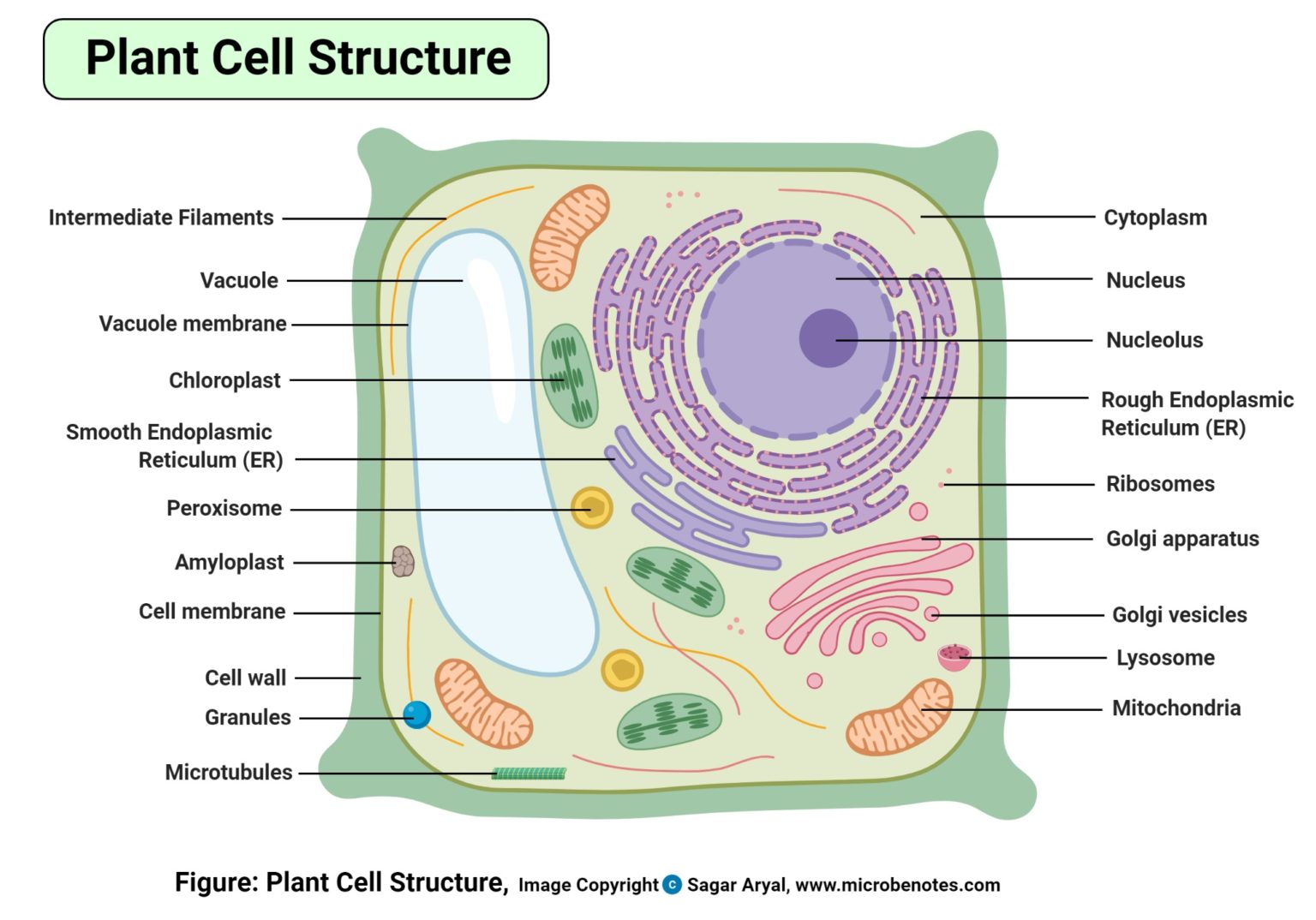
Plant Cell Definition Labeled Diagram Structure Parts Organelles from microbenotes.com The organelles of the organism and its cytoplasm are therefore bound by a plasma membrane that allows for easier movement. Electron microscope, microscope that attains extremely high resolution using an electron beam instead of a beam of light to illuminate the object of study. The explosion of knowledge brought about by improvements in microscopy, biochemistry, and genetics. Certain structures can be seen only under an electron microscope. Use the following animation to explore bacterial structure. The modern understanding of cellular substructure began with the use of the electron microscope. Just under the rigid cell wall is the more fluid cell membrane. Light passes from a bulb under the stage, through a …
Just under the rigid cell wall is the more fluid cell membrane.
Aug 20, 2018 · structure of cell the detailed structure of a cell has been studied under compound microscope and electron microscope. Electron microscope, microscope that attains extremely high resolution using an electron beam instead of a beam of light to illuminate the object of study. Fundamental research by many physicists in the first quarter of the 20th century suggested that cathode rays (i.e., electrons) might be used in some way to increase microscope resolution. The explosion of knowledge brought about by improvements in microscopy, biochemistry, and genetics. Let us have a detailed look at the plant cell, its structure and functions of different plant cell organelles. The plant cell is surrounded by a cell wall which is involved in providing shape to the plant cell. This means that divided further, its components (electrons, protons, and neutrons) do not retain the properties of the element. The modern understanding of cellular substructure began with the use of the electron microscope. Observation of euglena under more powerful electron microscopes have revealed the presence of an ornamented pellicle under the plasma membrane. Use the following animation to explore bacterial structure. Light passes from a bulb under the stage, through a … The structure of a cell as seen under an electron microscope is called ultrastructure. Unlike most plant cells, this species do not have a cell wall.
The organelles of the organism and its cytoplasm are therefore bound by a plasma membrane that allows for easier movement plant cell under electron microscope. The structure of a cell as seen under an electron microscope is called ultrastructure.
Share :
Post a Comment
for "Structure Of Plant Cell Under Electron Microscope / Cell And Organelles Dr Jastrow S Electron Microscopic Atlas : Electron microscope, microscope that attains extremely high resolution using an electron beam instead of a beam of light to illuminate the object of study."


Post a Comment for "Structure Of Plant Cell Under Electron Microscope / Cell And Organelles Dr Jastrow S Electron Microscopic Atlas : Electron microscope, microscope that attains extremely high resolution using an electron beam instead of a beam of light to illuminate the object of study."