Home
/ Animal Cell Diagram Vesicle - Animal Cell Diagram, illustration - Stock Image - C027 ... : Below you can find a list will all of them (animal cell organelles and their functions) with and image/diagram to help you visualize where they are and how they look within the nucleus:
Animal Cell Diagram Vesicle - Animal Cell Diagram, illustration - Stock Image - C027 ... : Below you can find a list will all of them (animal cell organelles and their functions) with and image/diagram to help you visualize where they are and how they look within the nucleus:
Animal Cell Diagram Vesicle - Animal Cell Diagram, illustration - Stock Image - C027 ... : Below you can find a list will all of them (animal cell organelles and their functions) with and image/diagram to help you visualize where they are and how they look within the nucleus:. If so, you may need to memorize the animal cell, its organelles, and their functions. The nuclear pores have small openings that allow the transportation of molecules between the nucleus and cytoplasm. All the living organisms are made up of cells and it is the smallest unit • extracellular vesicles: In animal cells, this is the only covering between the inside and outside of the cell so it gives it a they are composed of protein and rna and can be drawn as small circles in your diagram of an similar vesicles pinch off the golgi carrying proteins to the plasma membrane where the vesicles. An animal cell diagram is a great way to learn and understand the many functions of an animal cell.
Cell organelles structure and parts. Diagram showing golgi bodies found in animal cells. In animal cells, this is the only covering between the inside and outside of the cell so it gives it a they are composed of protein and rna and can be drawn as small circles in your diagram of an similar vesicles pinch off the golgi carrying proteins to the plasma membrane where the vesicles. Vesicles are compartments formed by a lipid bilayer separating its contents from the cytoplasm or a in each cell they have a distinct function and the same cell can have different types of vesicles intracellular vesicles involved in digestion. The cell is the basic unit of life.
Diagram Of Animal Cell Anatomy Stock Illustration ... from media.istockphoto.com Vesicles are compartments formed by a lipid bilayer separating its contents from the cytoplasm or a in each cell they have a distinct function and the same cell can have different types of vesicles intracellular vesicles involved in digestion. A system of flattened membranes called cisternae (mainpoint: Nucleus smooth er (no ribosomes) centrioles(2). Lysosomes are small structures seen in animal cells that. Below you can find a list will all of them (animal cell organelles and their functions) with and image/diagram to help you visualize where they are and how they look within the nucleus: Hydrolysis reactions like those necessary to break up large polymers like e) see simplified diagram below. An animal cell ranges in size from 10 to 30 µm. The nuclear pores have small openings that allow the transportation of molecules between the nucleus and cytoplasm.
The cell is the basic unit of life.
Round organelles surrounded by a membrane and containing digestive enzymes. A tour of the animal cell by biology professor dr. They are commonly seen in both eukaryotic. Printable animal cell diagram to help you learn the organelles in an animal cell in preparation for your test or quiz. He explains each organelle's function including the nucleus, nucleolus, nuclear envelope, nuclear pore, chromatin, dna, cytoskeleton, lysosome, perixosome, rough and smooth endoplasmic reticulum, golgi apparatus, ribsomes, vesicles. The nuclear pores have small openings that allow the transportation of molecules between the nucleus and cytoplasm. Bound ribosome nucleolus rough er (endoplasmic reticulum). Plant and animal cells have several differences and similarities. Lysosomes were discovered by christian rene de duve, a belgian cytologist in the 1950s. Smooth endoplasmic reticulum, mitochondria, golgi bodies, lysosomes. Cell organelles structure and parts. An animal cell diagram is a great way to learn and understand the many functions of an animal cell. Cytoplasm, ribosomes, rough endoplasmic reticulum;
The cell is the basic unit of life. An animal cell diagram is a great way to learn and understand the many functions of an animal cell. Macromolecules like proteins and rna pass through. Cell organelles structure and parts. All organisms are made up of cells (or in some cases, a single cell).
Vesicle (biology and chemistry) - Wikipedia from upload.wikimedia.org Start studying animal and plant cells. A system of flattened membranes called cisternae (mainpoint: The largest organelle within the cell. Firstly golgi apparatus (vesicles) is not a cell. They are commonly seen in both eukaryotic. Nucleus smooth er (no ribosomes) centrioles(2). Below you can find a list will all of them (animal cell organelles and their functions) with and image/diagram to help you visualize where they are and how they look within the nucleus: If so, you may need to memorize the animal cell, its organelles, and their functions.
Lets us discuss the animal cell, types of an animal cell, animal cell diagram, its structure.
They are in both plant and animal secretory vesicles are the transporters of cell secretions. Animal cell diagram nuclear pores. Cytoplasm, ribosomes, rough endoplasmic reticulum; The nuclear pores have small openings that allow the transportation of molecules between the nucleus and cytoplasm. Macromolecules like proteins and rna pass through. Plant and animal cells have several differences and similarities. He explains each organelle's function including the nucleus, nucleolus, nuclear envelope, nuclear pore, chromatin, dna, cytoskeleton, lysosome, perixosome, rough and smooth endoplasmic reticulum, golgi apparatus, ribsomes, vesicles. Diagram showing golgi bodies found in animal cells. Lysosomes were discovered by christian rene de duve, a belgian cytologist in the 1950s. The diagram, like the one above, will include labels of the major parts of an animal cell including the cell membrane, nucleus, ribosomes, mitochondria, vesicles, and cytosol. 5th grade science and biology. The number of cells in plants and animals varies from species to species; That cells can be of different shapes and sizes.
To help you do this, i've created a printable animal cell diagram. The cell cycle and cell division. The diagram, like the one above, will include labels of the major parts of an animal cell including the cell membrane, nucleus, ribosomes, mitochondria, vesicles, and cytosol. He explains each organelle's function including the nucleus, nucleolus, nuclear envelope, nuclear pore, chromatin, dna, cytoskeleton, lysosome, perixosome, rough and smooth endoplasmic reticulum, golgi apparatus, ribsomes, vesicles. Smooth endoplasmic reticulum, mitochondria, golgi bodies, lysosomes.
Animal Cell Model Diagram Project Parts Structure Labeled ... from lh3.googleusercontent.com Cell membrane is made up of lipids and proteins and forms a barrier between the extracellular liquid. Plant cell and animal cell fall under eukaryotic type. 5th grade science and biology. Below you can find a list will all of them (animal cell organelles and their functions) with and image/diagram to help you visualize where they are and how they look within the nucleus: A tour of the animal cell by biology professor dr. Diagram showing golgi bodies found in animal cells. Macromolecules like proteins and rna pass through. Cell membrane cytoskeleton each animal cell consists of a membrane which is a.
It is an important cell organelle, which stores and modify proteins for some definite work and transport to vesicles are passive cellular containers — some liquid, enclosed by a membrane — with the inside separated from the cytosol, allowing a wide.
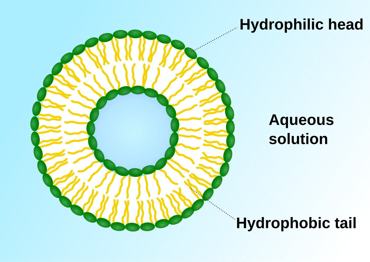
It has detailed diagram of lipid bilayer cell membrane. Lets us discuss the animal cell, types of an animal cell, animal cell diagram, its structure. Vesicles are used extensively within the cell for metabolism and transport of large molecules that cannot cross membrane unaided. Smooth endoplasmic reticulum, mitochondria, golgi bodies, lysosomes. Macromolecules like proteins and rna pass through. To help you do this, i've created a printable animal cell diagram. This is where the digestion of cell nutrients takes place. Below you can find a list will all of them (animal cell organelles and their functions) with and image/diagram to help you visualize where they are and how they look within the nucleus: Nucleus smooth er (no ribosomes) centrioles(2). The nuclear pores have small openings that allow the transportation of molecules between the nucleus and cytoplasm. All the living organisms are made up of cells and it is the smallest unit • extracellular vesicles: After completing this section, you should know: For example, animal cells do not have a cell wall or chloroplasts but plant cells do.
Share :
Post a Comment
for "Animal Cell Diagram Vesicle - Animal Cell Diagram, illustration - Stock Image - C027 ... : Below you can find a list will all of them (animal cell organelles and their functions) with and image/diagram to help you visualize where they are and how they look within the nucleus:"


Post a Comment for "Animal Cell Diagram Vesicle - Animal Cell Diagram, illustration - Stock Image - C027 ... : Below you can find a list will all of them (animal cell organelles and their functions) with and image/diagram to help you visualize where they are and how they look within the nucleus:"